Observation Notes
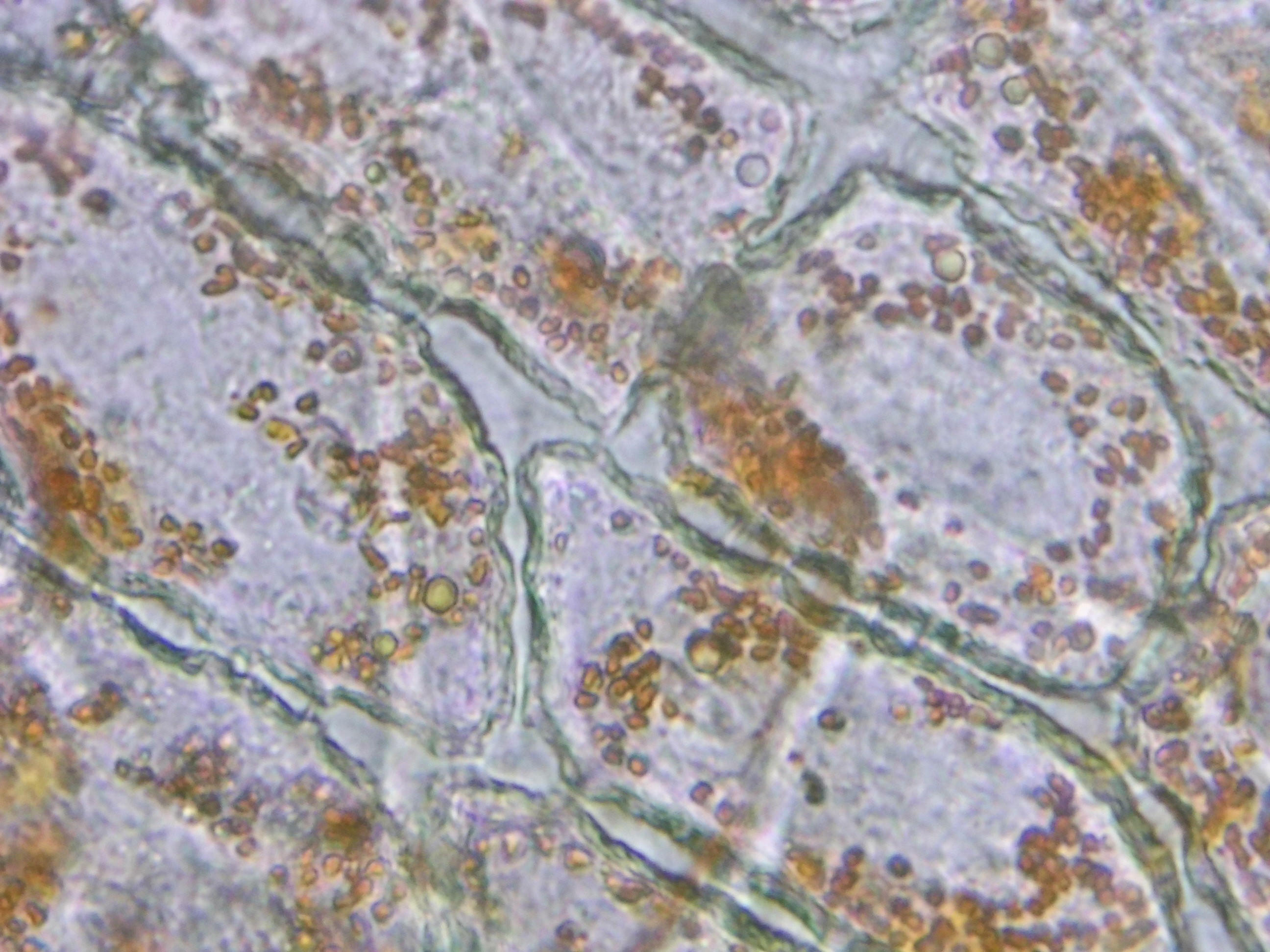
This cross section was prepared using a standard microtome at 10μm thickness and stained with toluidine blue. The vascular bundles are clearly visible, showing both xylem and phloem tissue arrangements.
Key Structures
- Upper epidermis — single layer of tightly packed cells with a waxy cuticle
- Palisade mesophyll — columnar cells rich in chloroplasts, primary site of photosynthesis
- Spongy mesophyll — irregularly shaped cells with large air spaces for gas exchange
- Vascular bundle — central vein containing xylem (water transport) and phloem (sugar transport)
- Lower epidermis — contains stomata for CO₂ intake and water vapor release
Staining Details
Toluidine blue O is a metachromatic dye, meaning it produces different colors depending on the tissue it binds to. Lignified cell walls (xylem) stain blue-green, while non-lignified walls (phloem, mesophyll) stain purple-pink. This differential staining makes it easy to distinguish tissue types at a glance.